Diagram Of Shoulder And Arm / Osteoarthritis Oa Of The Elbow And Shoulder : The bones of the shoulder are the humerus (the upper arm bone), the scapula (the shoulder blade), and the clavicle (the collar bone).
Diagram Of Shoulder And Arm / Osteoarthritis Oa Of The Elbow And Shoulder : The bones of the shoulder are the humerus (the upper arm bone), the scapula (the shoulder blade), and the clavicle (the collar bone).. Muscles of the shoulder : It rotates the forearm and also flexes the elbow. The shoulder joint is formed where the humerus (upper arm bone) fits into the scapula (shoulder blade), like a ball and. Diagramme schnell und einfach erstellen. The shoulder is one of the largest and most complex joints in the body.
Muscles of the shoulder : The scapula is also called the shoulder. We hope this picture right arm muscle and tendon anatomy can help you study and research. This holds your upper arm bone to your shoulder blade and helps you rotate your arm, hold it straight out and lower it. The main shoulder joint, called the glenohumeral joint, is formed
Muscles Of The Shoulder Upper Arm Diagram Quizlet from o.quizlet.com The etiology is most of the time traumatic and related either to sport or accidents. The rotator cuff muscles are important stabilizers and movers of the shoulder joint. Shoulder joint injuries can be head. The three bones of the shoulder are the: Four of them are found on the anterior aspect of the shoulder, whereas the rest are located on the shoulder's posterior aspect and in the back. For more anatomy content please follow us and visit our website: The shoulder plays a key role in the blood flow to the arms. The roof of the shoulder is formedby a part of the scapula called the acromion.
The bones of the shoulder are:
The shoulder joint is formed where the humerus (upper arm bone) fits into the scapula (shoulder blade), like a ball and. Diagramme schnell und einfach erstellen. Is the wear and tear of shoulder cartilage until bare bone is exposed. Test your knowledge of the clavicle, scapula and humerus with our labeled diagram exercises and quizzes! Your collarbone (clavicle), head of the arm bone (humerus) and actual shoulder bade (scapula). We hope this picture right arm muscle and tendon anatomy can help you study and research. Movement in this part of the body is more each time the arm is raised, not only does the ball of the humerus move in the socket of the glenoid, but the clavicle and the acromion rotate 40 degrees. There are two joints within the shoulder that can be affected by osteoarthritis. It rotates the forearm and also flexes the elbow. We think this is the most useful anatomy picture that you need. The bones of the upper arm include the: A final test for frozen shoulder is to stand with both arms at the sides and the elbows flexed at ninety degrees. Four of them are found on the anterior aspect of the shoulder, whereas the rest are located on the shoulder's posterior aspect and in the back.
The bicep has a total of two shoulder tendons. Shoulder joint injuries can be head. Where the rounded top of the arm bone (humerus) contacts the shoulder blade is. The glenoid is covered with smooth cartilage. See more ideas about muscle anatomy, anatomy, shoulder muscle anatomy.
Human Arm Anatomical Structure Download Scientific Diagram from www.researchgate.net There are actually four joints that make up the shoulder. This is called the glenoid. In this episode of eorthopodtv, orthopaedic surgeon randale c. Three of them are located in the anterior compartment — the biceps brachii, brachialis, and coracobrachialis, while the forth is located in the posterior compartment — the triceps brachii). Anatomynote.com found right arm muscle and tendon anatomy from plenty of anatomical pictures on the internet. Diagram of the shoulder, including the location of the rotator cuff. Movement in this part of the body is more each time the arm is raised, not only does the ball of the humerus move in the socket of the glenoid, but the clavicle and the acromion rotate 40 degrees. The shoulder is one of the largest and most complex joints in the body.
The humerus is the (upper) arm bone.
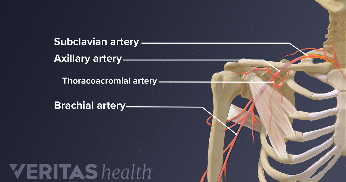
17 photos of the diagram of shoulder muscles and tendons. It joins with the scapula above at the shoulder joint (or glenohumeral joint) and with the ulna and radius below at the elbow joint. The bicep has a total of two shoulder tendons. Shoulder pain is one of the most common complaints in the outpatient setting. Where the rounded top of the arm bone (humerus) contacts the shoulder blade is. The humerus is the (upper) arm bone. Shoulder joint injuries can be head. The largest bone of the arm, the humerus connects to the scapula and clavicle in the shoulder. Arm science bone anchor chart shoulder human skeleton diagram health human body. The armpit and shoulder serve as the meeting place for the torso and arms, so major vessels close to the heart travel through these areas. Your collarbone (clavicle), head of the arm bone (humerus) and actual shoulder bade (scapula). Flexibility and joint limitations : The roof of the shoulder is formedby a part of the scapula called the acromion.
From the arm muscle diagram above, the muscles of the arm that can be seen easily on the surface include biceps, triceps, brachioradialis, extensor carpi radialis longus, and deltoid.biceps are large muscle of the upper arm is formally known as the biceps brachii muscle, and rests on top of the humerus bone. There are four muscles in you upper arm, which is delimited by your shoulder joint and your elbow joint. The upper arm includes the shoulder as well as the area between the shoulder and elbow joint. Three of them are located in the anterior compartment — the biceps brachii, brachialis, and coracobrachialis, while the forth is located in the posterior compartment — the triceps brachii). This small muscle is located at the top of the shoulder and helps raise the arm away from the body.
Blood And Nerve Supply Of The Shoulder from embed.widencdn.net The shoulder joint is composed of the glenoid (the shallow shoulder socket) and the head of the upper arm bone known as the humerus (the ball). Shoulder and arm (labeled) 1 of 1 shoulder diagram human body health human bone arm anchor chart skeleton science. In this episode of eorthopodtv, orthopaedic surgeon randale c. This is called the glenoid. Diagram of shoulder and arm : The bones of the shoulder are the humerus (the upper arm bone), the scapula (the shoulder blade), and the clavicle (the collar bone). The roof of the shoulder is formedby a part of the scapula called the acromion. We hope this picture right arm muscle and tendon anatomy can help you study and research.
The three bones of the shoulder are the:
The shoulder is one of the largest and most complex joints in the body. Where the rounded top of the arm bone (humerus) contacts the shoulder blade is. The bones of the shoulder are: The shoulder joint can sometimes become narrowed and arthritic, and spurs can form on the undersurface. The upper arm includes the shoulder as well as the area between the shoulder and elbow joint. We hope this picture right arm muscle and tendon anatomy can help you study and research. Sechrest, md narrates an animated tutorial on the basic anatomy of the shoulder. From the arm muscle diagram above, the muscles of the arm that can be seen easily on the surface include biceps, triceps, brachioradialis, extensor carpi radialis longus, and deltoid.biceps are large muscle of the upper arm is formally known as the biceps brachii muscle, and rests on top of the humerus bone. The bones of the shoulder are the humerus (the upper arm bone), the scapula (the shoulder blade), and the clavicle (the collar bone). For more anatomy content please follow us and visit our website: It joins with the scapula above at the shoulder joint (or glenohumeral joint) and with the ulna and radius below at the elbow joint. The roof of the shoulder is formedby a part of the scapula called the acromion. Injuries to the rotator cuff are common, but treatment is often successful.
A final test for frozen shoulder is to stand with both arms at the sides and the elbows flexed at ninety degrees diagram of shoulder. What are common rotator cuff injuries?


0 Komentar